BOISE, Idaho — This story originally appear in the Idaho Press.
For years, the medical community has known high breast density poses a higher risk for developing breast cancer, but in September that knowledge was finally recognized through a new ruling.
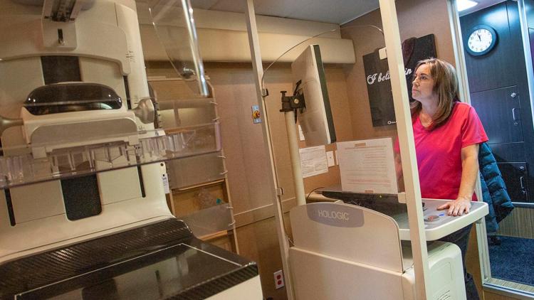
The U.S. Food and Drug Administration now requires all mammography facilities to include a breast density assessment in a patient’s medical report after a mammogram. While this might not be a big change for Idaho mammogram reports, the ruling brings a new level of standardization to the medical community and the reason is clear: breast density matters.
October is Breast Cancer Awareness Month, meant to spread awareness of victims and survivors of breast cancer.
According to the National Breast Cancer Foundation, in the U.S., one in eight women will be diagnosed with breast cancer in their lifetime. In 2024, around 310,720 women and 2,800 men will be diagnosed with invasive breast cancer.
Breasts are typically composed of two types of tissue — fatty and glandular. But the compositions on everyone is a little different. Some women have more fatty than glandular tissue, while other women have more glandular than fatty.
Women who have more glandular tissue have denser breasts.
According to breast cancer surgeon Elizabeth Prier, alerting patients of their breast density after a mammogram is not new to Saint Alphonsus because it is one factor that can make a patient at risk for developing breast cancer. Other factors can include family history and age, Prier said.
“The risk of breast cancer increases as you age,” Laura Linstroth, director of breast imaging for Saint Alphonsus, said. “It’s always going to increase with time. It never really plateaus with a certain age or goes down, so by the time women are in their 80s to 90s, we estimate that their risk of breast cancer is about one in eight women.”
Women with dense breast tissue have an increased risk for developing breast cancer for two reasons: one, because glandular tissue (dense breast tissue) has more of a risk for cancer than fatty tissue does; and two, because mammograms on women with dense tissue don’t detect breast cancer until it’s developed into a larger mass.
In other words, women with more glandular breast tissue are more likely to have breast cancer that goes unnoticed, making future treatment more strenuous. Those women benefit from secondary screenings, specifically whole breast ultrasounds and abbreviated screening MRI, Prier said. Early detection is pivotal, which is why women should know about their breast density, so they can get extra screenings if necessary.
A whole breast ultrasound gets imaging of the whole breast via a screening technique. It’s a simple, painless procedure with no radiation or IVs, according to Linstroth.
An abbreviated breast MRI screening is a six-minute test that uses contrast through an IV to get images of the breast. According to Prier, it’s a good way to look for smaller tumors and is the most sensitive supplemental screening to a mammogram at detecting small cancers.
“One of the things that’s important for women to understand is that these alternative options for screening aren’t in place of a mammogram,” Prier said. “They’re complementary. There are things that a mammogram sees, even if you have dense breast tissue, that these other technologies, like whole breast ultrasound or breast MRI, might not see.”
For example, precancerous changes tend to show up on mammograms, not whole breast ultrasounds or MRIs, Prier said.
“If your letter says that you have increased breast density and need to consider supplemental screening,” Prier said, “women should consider this automated whole breast ultrasound or abbreviated breast MRI. We offer both of those modalities at Intermountain Medical Imaging at our Meridian campus.”
The sooner the cancer is caught, the better a patient’s chances of survival are. And the earlier the cancer stage is, the less treatment is required, Prier said.
“If we catch cancer when it’s stage three, you’re going to probably need chemotherapy, surgery, radiation. If we catch a cancer at a stage one, it could just be surgery and radiation,” Prier said.
Abbreviated breast MRIs are only available at Intermountain Medical Imaging in Meridian, Linstroth said. There are several locations in Idaho where women can get an automated whole breast ultrasound, but all of them have popped up in the last year.
“It’s really exciting,” Linstroth said. “We have all of this new technology now available to a widespread population.”
The thing that’s missing right now is universal insurance coverage, but according to Linstroth, laws typically precede insurance coverage.
“It’s going to be a barrier that will hopefully improve over time,” she said. “We don’t want insurance to be a barrier to care.”
For now though, it may be for some.
Mammograms remain the golden standard for women, who should start getting mammograms every year beginning at the age of 40.
Around the age of 30, women should begin talking to their health care provider about their risk factors for breast cancer, Linstroth said.
“If they’re feeling any new lump or bump, having clear bloody nipple discharge or pain in one spot in the breast that doesn’t go away and is not related to their menstrual cycle or muscular injury, those are things that we like to see women in for an ultrasound if they’re younger, and an ultrasound and a mammogram if they’re usually 30 and over,” Linstroth said.
Most breast cancers that are detected by mammogram or other imaging are stage one cancers, which typically have a 90 to 95% survival and cure rate, Linstroth said.
“It’s not the dire circumstance that it was many years ago,” Linstroth said. “Now, we can pretty much cure most breast cancers and women can go on to be sisters, moms, daughters, grandmas, and healthy women in the workforce. Everything that they want to do, they can still do even with the diagnosis of breast cancer.”
This article originally appeared in the Idaho Press, read more on IdahoPress.com.


